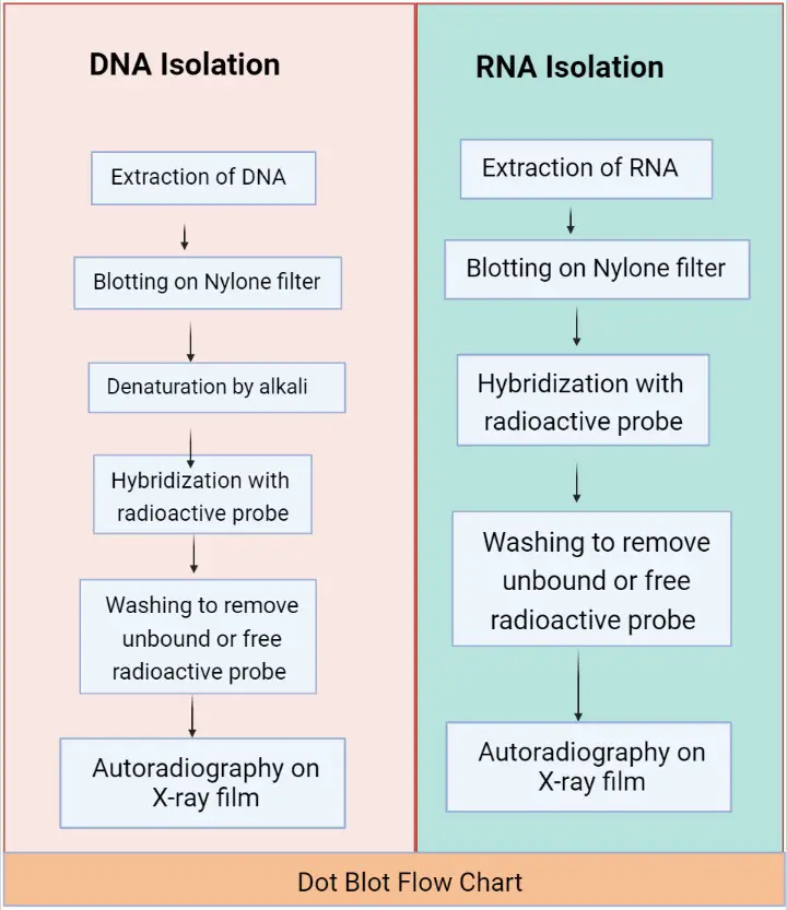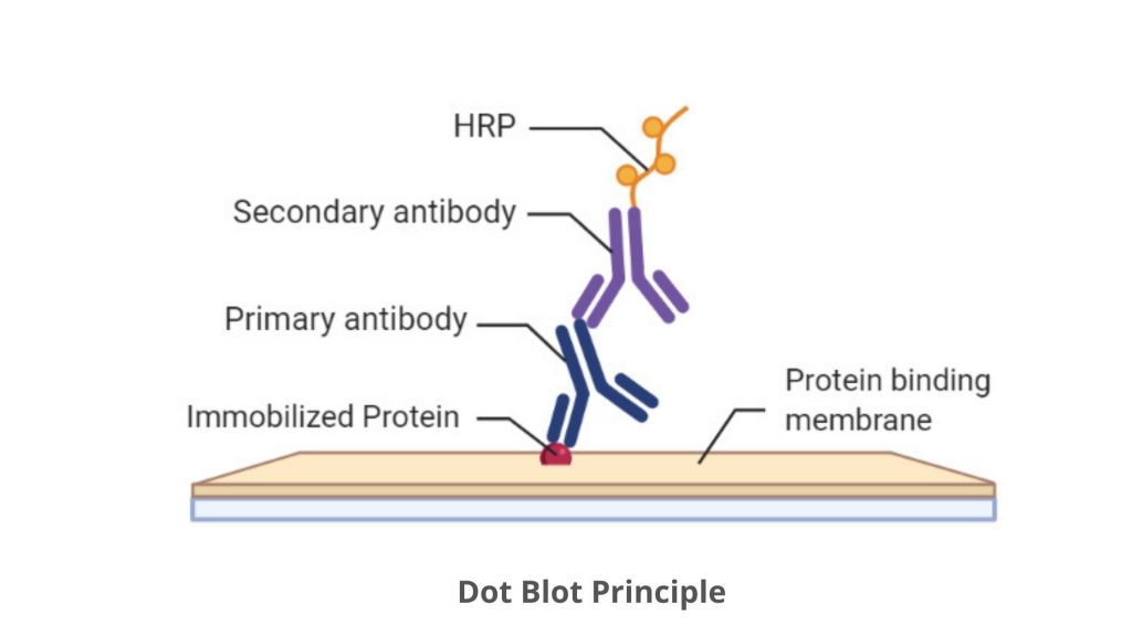

In the first set of experiments, we used leukocyte DNA donated by 28 young and middle-age adults. Based on dot blot analysis, the method requires little amount of DNA (∼20 ng/assay), and moreover is inexpensive and easy to perform. This need will only escalate with the anticipated expansions of research and clinical applications that will require telomere length measurements. For epidemiological research, the TRF length analysis and qPCR method are typically preferred over the other methods, but they too display considerable problems that limit their practical use.Ĭlearly, there is a growing need for a simple, specific and sufficiently precise method to measure telomere length for research and clinical purposes. Although these techniques have been successfully employed in broadening understanding the roles of telomeres in human health and disease, they are also fraught with shortcomings ( 20, 21 ) that have contributed to a growing debate about their research applications and usefulness in clinical settings. Methods to measure telomere length/content include Southern blot analysis of the terminal restriction fragment (TRF) lengths ( 1 ), quantitative PCR (qPCR) ( 12, 13 ), fluorescent in situ hybridization (FISH) ( 14, 15 ), slot blot or dot blot analysis ( 16–18 ) and single telomere length analysis (STELA) ( 19 ). Moreover, shortened LTL is often expressed in various forms of aplastic anemia ( 11 ).

Relatively short LTL has also been found to be associated with diminished survival of elderly persons ( 8–10 ). These studies have established that LTL is inversely related to age and is relatively short in individuals who suffer from aging-related diseases, principally atherosclerosis ( 5–7 ). Accordingly, telomere dynamics (telomere length and its shortening) have been the focus of intense interest in both clinical and epidemiological studies, which have primarily measured leukocyte telomere length (LTL). Cellular replication figures in mammalian development, tissue maintenance and in aging it is also a key player in the runaway cellular replication that is the hallmark of cancer ( 3, 4 ). In these cells, telomere length is not only a record of cell replication but it is also an index of replicative potential. Telomeres, the TTAGGG repeats at the ends of chromosomes, undergo progressive shortening in replicating cells that lack active telomerase-the reverse transcriptase that adds telomere repeats to the end of the chromosomes ( 1, 2 ). The method might help researchers and clinicians alike in understanding risks for and extent of human diseases.

The method is also simple to use, has a relatively low interassay coefficient of variation (<6%), retains its precision in moderately degraded DNA and can be forged for high throughput analysis. Moreover, the T/Dx data are highly correlated linearly with mean telomere lengths derived from Southern blots of the terminal restriction fragments ( r > 0.96, P < 0.0001). The method requires ∼20 ng of DNA per assay. The T normalized for Dx (T/Dx) of each dot is a measure of telomere content. In each dot, the DNA content is measured by a DNA stain (Dx) and the telomeric DNA content is measured with a telomeric probe (T). Here, we describe a novel method to measure the relative telomere DNA content by dot blot analysis. However, present methods to measure telomere length are fraught with shortcomings that limit their use. Telomeres play a central role in human cancer, cardiovascular aging and possibly longevity.


 0 kommentar(er)
